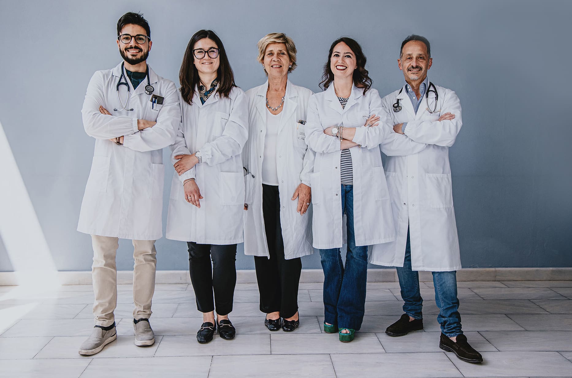
Arithmology Center
The San Rossore Arrhythmology Center is a unit specializing in the treatment of arrhythmias that aims to offer effective solutions to restore and maintain a normal heart rhythm.

What are arrhythmias:
Cardiac arrhythmias are alterations in the rhythm of the heart, which can manifest as irregular heartbeats that are too fast or too slow. These abnormalities can be caused by several factors, including structural heart disorders, electrolyte imbalances, electrical conduction problems or other medical conditions.
Cardiac arrhythmias can affect the frequency, rhythm, or regularity of heart contractions. In fact, the heart beats normally due to an electrical excitation-conduction system that generates coordinated electrical impulses. These impulses control the rhythm and sequence of contractions of the heart chambers (atria and ventricles), enabling the heart to pump blood effectively.
However, in people with arrhythmias, the heart’s electrical excitation-conduction system may be disturbed, causing abnormal rhythms. Arrhythmias can be classified in several ways, but the two main categories are bradyarrhythmias (slow heartbeat) and tachyarrhythmias (rapid heartbeat).
The bradyarrhythmias include sinus bradycardias and heart blocks, in which electrical impulses are generated or conducted in a slowed manner causing the heart rate to be lower than normal. This can lead to symptoms such as fatigue, fainting or dizziness.
The tachyarrhythmias include supraventricular tachycardias, atrial flutter and fibrillation, ventricular arrhythmias (ventricular tachycardia and ventricular fibrillation). Supraventricular tachycardia is a condition in which an accelerated heart rhythm occurs due to abnormal electrical impulses originating above the ventricles. Atrial fibrillation is one of the most common arrhythmias, characterized by an irregular and fast rhythm in the upper chambers of the heart (atria). Complex ventricular arrhythmias are potentially dangerous arrhythmias in which the heart rhythm can become chaotic and very fast, causing a loss of efficiency in the heart pump.
The causes of cardiac arrhythmias can vary and include preexisting heart diseases such as hypertension, ischemic heart disease, and congenital malformations but can also be caused by electrolyte imbalances, thyroid dysfunction, use of specific drugs, substance abuse, stimulants or alcohol, genetic factors, stress, and anxiety.
The treatment of arrhythmias depends on their severity, associated symptoms and the patient’s health condition. Treatment options may include lifestyle modifications, antiarrhythmic drugs, radiofrequency ablation or cryotherapy, implantation of devices such as pacemakers or implantable cardioverter defibrillators (ICDs), and in some cases, surgery
The diagnosis and treatment of arrhythmias is carried out by Cardiologists who specialize in the management of Cardiac Arrhythmias. The San Rossore Arrhythmology Center is a unit specializing in the treatment of these conditions and aims to offer effective solutions to restore and maintain a normal heart rhythm.
In the Arrhythmology Center, a team of experienced cardiologists focuses on the diagnosis and evaluation of cardiac arrhythmias. Patients undergo a series of specific examinations, such as electrocardiogram (ECG), which records the heart’s electrical activity, echocardiogram, cardiac MRI, and stress test, which are used to identify any underlying structural heart disease, and, last but not least, Holter monitoring to record cardiac activity. As instrumental documentation of arrhythmia remains the key moment of diagnosis, in recent years, new long-term electrocardiographic monitoring systems have been developed and introduced (>24h), which revolutionized the diagnostic pathway of cardiac arrhythmias.
These devices are useful for detecting and recording the heart’s electrical activity for prolonged periods, enabling physicians to obtain a more complete and detailed view of the patient’s heart rhythm, along with related symptomatic correlations. Some of the major long-term electrocardiographic monitoring systems include:
48-72-hour Holter ECG:
The Holter ECG is a portable device that continuously records the electrical activity of the heart over a period of 48-72 hours. The patient wears small electrodes on the chest, which are connected to a portable recorder. During the monitoring period, the patient can carry out normal daily activities. At the end of the recording period, the device is returned to the medical center, where the data are analyzed for arrhythmias or heart rhythm abnormalities.
External Event Recorder:
“External event recorders” are handheld devices that the patient can activate manually when experiencing symptoms or episodes of arrhythmia. When a symptom occurs, the patient presses a button on the device, which records the heart’s electrical activity for a short period, usually several minutes to several hours. These devices are useful for capturing sporadic events or transient symptoms that might not be recorded by other monitoring modalities. The same recording can be made with a portable ECG with durations typically as short as 30 seconds, which, via special apps can be emailed as a pdf to the referring physician for timely diagnosis.
External Loop Recorder:
“External loop recorders” are devices similar to “event recorders,” but are capable of continuously recording the heart’s electrical activity for a longer period of time, often up to 30 days. These devices are used to monitor patients with less frequent arrhythmias or intermittent symptoms.
Internal Loop Recorder:
“Internal loop recorders,” also known as implantable loop recorders, are smaller devices that are surgically implanted into the subcutaneous tissue. These devices can continuously monitor the heart’s electrical activity for longer periods of time than external loop recorders, often up to 3 years or more. Data can be transmitted to physicians remotely via wireless connection.
The use of these new long-term monitoring systems has enabled physicians to more accurately diagnose and treat arrhythmias, improving the clinical management of patients and enabling more personalized treatment pathways. These devices are particularly useful for patients with intermittent symptoms or arrhythmias that are difficult to detect with conventional diagnostic techniques.
Finally, in selected cases in which the clinical diagnosis remains uncertain, additional invasive tests, such as endocavitary electrophysiological study, may be necessary to obtain an accurate diagnosis and plan the appropriate treatment.
Correct electrocardiographic documentation of clinical arrhythmia is critical to planning appropriate treatment. The San Rossore Arrhythmology Center is a unit specializing in ultraspecialty diagnostics of cardiac arrhythmias.
The Arrhythmology Center offers a wide range of pathways and therapeutic treatments for cardiac arrhythmias. After a comprehensive evaluation, the team of specialists develops an individualized treatment plan for each patient, taking into account the severity of arrhythmias, symptoms and individual health conditions. Available treatment options include:
Drug therapy: specific drugs are prescribed to control and regulate heart rhythm, thereby reducing the frequency and intensity of arrhythmias.
Electrical cardioversion: a procedure in which a controlled electrical discharge is applied to the heart to restore a normal heart rhythm.
Catheter ablation: a minimally invasive procedure in which one or more catheters are inserted into veins or arteries and guided to the heart to destroy the cells responsible for arrhythmia.
Implantation of cardiac devices: In some cases, it may be necessary to implant a pacemaker or cardiac defibrillator to monitor and correct the heart rhythm.
Regarding implantable electrical devices, alongside traditional “transvenous” (using leads introduced through a central vein such as the subclavian), new technologies have been introduced in recent years, such as “leadless” pacemakers and subcutaneous defibrillators.
The pacemaker leadless are implantable devices that are placed directly into the heart, without the need for traditional electrodes (leads). These miniaturized, capsule-sized devices are inserted through a minimally invasive procedure, usually through a catheter that is inserted into the femoral vein and guided to the heart. The absence of traditional leads minimizes the risk of complications associated with such devices, such as infection or catheter malfunction.
The Subcutaneous defibrillators are implantable devices designed to monitor the electrical activity of the heart and provide an electrical discharge when a severe arrhythmia, such as ventricular fibrillation, is detected. Unlike conventional defibrillators, which are implanted internally and require electrodes passing through the veins to the heart, subcutaneous defibrillators are placed entirely under the patient’s skin, in the left lateral region of the chest. This approach reduces the risk of complications associated with intravascular electrodes and simplifies the implantation procedure.
All of these devices, which are regularly available and implanted at the Casa di Cura San Rossore, represent important innovations in the treatment of cardiac arrhythmias, offering safer and more convenient solutions for patients. Of course, the choice of the most appropriate therapeutic device will depend on the assessment of the medical specialist, taking into account the patient’s individual characteristics and specific clinical needs.
As part of the radiofrequency ablation for the treatment of cardiac arrhythmias, electroanatomical mapping systems and zero-beam techniques have revolutionized the therapeutic approach, providing physicians with more precise and effective tools to identify and treat the areas responsible for arrhythmias.
The electroanatomical mapping systems allow physicians to obtain a detailed three-dimensional representation of the heart and its electrical structures during the ablation procedure and to create real-time maps of the heart’s electrical activities. During ablation, physicians can use these maps to accurately identify the problem areas responsible for arrhythmias and guide catheters in a targeted manner to apply radiofrequency to the areas to be treated. The use of electroanatomic mapping systems has greatly improved the accuracy and effectiveness of ablations, reducing the risk of complications, increasing the success rate of treatments, and
minimizing traditional radiological exposure during the procedure. Indeed, the adoption of zero-ray techniques has helped to improve the safety of surgeries and reduce X-ray exposure for patients and operators.
The combined use of electroanatomic mapping systems and zero-ray techniques is a priority of the San Rossore Clinic. These approaches enable clinicians to more accurately identify the sources of arrhythmias, optimize the ablation strategy, and achieve a higher rate of therapeutic success, while ensuring maximum safety for patients and operators.
What are the most common symptoms of cardiac arrhythmias?
Symptoms of cardiac arrhythmias can vary from person to person, but the most common include palpitations (feeling of an irregular or accelerated heartbeat), feeling of the heart missing or stopping, dizziness, fatigue, dyspnea (difficulty breathing) and fainting. However, some arrhythmias may be asymptomatic and are often discovered during routine examinations.
What are the main causes of arrhythmias?
Cardiac arrhythmias can be caused by a variety of factors, including structural heart disease, such as ischemic heart disease, heart failure or cardiomyopathy, electrolyte imbalances (such as low potassium or magnesium levels), thyroid dysfunction, drug or alcohol abuse, excessive caffeine consumption, stress and anxiety. In some cases, arrhythmias may be hereditary.
Are cardiac arrhythmias dangerous?
The severity of cardiac arrhythmias varies greatly. Some arrhythmias are harmless and require no treatment, while others can be dangerous and require immediate intervention. It is important to consult a physician for accurate evaluation and proper management of arrhythmias. Some arrhythmias can increase the risk of complications, such as heart failure or stroke, if not treated properly.
What are the risk factors for the development of arrhythmias?
Risk factors for the development of cardiac arrhythmias include advanced age, the presence of pre-existing heart disease, such as hypertension, diabetes, obesity, smoking, alcohol or drug abuse, stress and anxiety, a family history of cardiac arrhythmias or inherited heart disease, imbalanced electrolytes, and taking certain medications.
How can I prevent cardiac arrhythmias?
Although it is not always possible to completely prevent cardiac arrhythmias, some measures can help reduce the risk of developing them. These include adopting a healthy lifestyle, such as eating a balanced diet, exercising regularly, avoiding smoking and limiting alcohol and caffeine consumption. It is also important to manage stress effectively and control underlying health conditions, such as hypertension or diabetes, through proper medical care.
What is an event recorder?
The event recorder is a portable device that records the electrical activity of the heart for extended periods, usually several weeks to several months.
What is an event recorder for?
The event recorder is useful for capturing occasional episodes of arrhythmias that might go undetected during brief monitoring. Patients can manually activate the event recorder when they experience symptoms of arrhythmia, allowing physicians to analyze the recorded events and make an accurate diagnosis.
What is the Portable ECG?
It is a portable medical device designed to monitor the electrical activity of the heart. It is a type of single- or multi-lead ECG (electrocardiogram) that allows users to record their heart activity via a mobile application.
How does the Portable ECG work?
The Portable ECG works by connecting the device to a phone or tablet via a sensor or electrode. The user holds the device in the hands or rests it on the chest for a few seconds, and records the electrical activity of the heart. The data are then displayed on the mobile application and can be shared with a health professional for analysis.
Why should I use the Portable ECG?
The Portable ECG can be used to record the ECG of the heart easily and conveniently in a wide variety of situations, especially on occasions of suspicious symptoms (e.g., heart palpitation) that are rare and of short duration, such that recording by traditional instruments (e.g., ECg op ECG Holter-24h) is not possible . It can be useful to monitor the electrical activity of the heart at home and to check the effectiveness of certain cardiac treatments. The device enables users to have more control over their heart health and share data with health professionals for accurate assessment.
Can I share the recorded data with my doctor?
Answer: Yes, the data recorded with can be shared with your doctor or a health professional. Kardia’s mobile application allows reports to be generated that can be sent via e-mail or other means of communication. Sharing data with a physician can provide important information for accurate assessment of cardiac health.
What is a loop recorder?
The loop recorder is a subcutaneous implantable device that continuously records the electrical activity of the heart for an extended period of time.
What is a loop recorder for?
The loop recorder is particularly useful for patients who present with sporadic episodes or unclear symptoms of arrhythmia. The device continuously records electrical activity, enabling physicians to detect and analyze arrhythmic events over time for accurate diagnosis.
What is an electrophysiological (EP) study?
Electrophysiological study is an invasive procedure in which catheters are inserted through veins to record the electrical activity of the heart and stimulate specific cardiac regions.
What is the purpose of an electrophysiological study?
Electrophysiological study is useful for identifying the origin and cause of arrhythmias, assessing response to antiarrhythmics, and guiding therapeutic decisions, such as radiofrequency ablation.
What is a subcutaneous ICD?
The subcutaneous ICD is an implantable device that continuously monitors heart rhythm and provides an electrical discharge to interrupt dangerous arrhythmias such as ventricular fibrillation.
What are the indications for subcutaneous ICD implantation?
Implantation of a subcutaneous ICD is recommended for patients at high risk of life-threatening ventricular arrhythmias, such as those with a history of sudden cardiac arrest or impaired ventricular function.
What is a leadless pacemaker?
The leadless pacemaker is an implantable device that stimulates the heart to maintain a regular heart rhythm, without the need for traditional (lead) leads.
What is ablation of paroxysmal supraventricular tachycardias?
Ablation of paroxysmal supraventricular tachycardias is a minimally invasive procedure that aims to disrupt the abnormal circuits in the heart’s electrical conduction system responsible for the tachycardias.
How is ablation of paroxysmal supraventricular tachycardias performed?
During ablation, catheters are inserted into the vascular system and guided to the heart. Using dedicated energy, an injury is created or the tissue that causes tachycardias is destroyed. This prevents the passage of abnormal electrical impulses and restores normal heart rhythm.
What are the benefits of ablation of paroxysmal supraventricular tachycardias?
Ablation can reduce or completely eliminate episodes of tachycardia, thus improving the patient’s quality of life. In addition, it can reduce or eliminate the need for long-term antiarrhythmic drugs.
What is atrial fibrillation ablation?
Atrial fibrillation ablation is a procedure that aims to interrupt or control the chaotic electrical activity in the upper chambers of the heart (atria) responsible for atrial fibrillation.
How is atrial fibrillation ablation performed?
During ablation, catheters are inserted into the vascular system and guided to the heart. Through the use of dedicated energy, an injury is created or heart tissue is blocked that contributes to atrial fibrillation. This helps restore a regular heart rhythm.
What are the benefits of atrial fibrillation ablation?
Ablation can significantly reduce the frequency and intensity of atrial fibrillation episodes, improving the patient’s quality of life. In some cases, it can completely eliminate the need for antiarrhythmic drugs. Ablation may also reduce the risk of stroke associated with uncontrolled atrial fibrillation.
Please note that these answers are general and each individual may have unique situations. It is always advisable to consult a medical specialist for personal evaluation
Head of Interventional Cardiology
Dr. Maria Grazia Bongiorni
Interventional Arithmology
Dr. Andrea Di Cori
Dr. Antonio Canu
Dr. Mario Giannotti
Dr. Matteo Parollo
Clinical Arithmology
Dr. Valentina Barletta
Dr. Sara Sbragi
INFO POINT
REQUEST INFORMATION
For questions, clarifications or other Info please fill out the form below, you will be contacted by our dedicated staff as soon as possible.


