Diagnostic Examinations
WORK IN PROGRESS
Page under construction
For information contact +39 050 586217 or send e-mail to info@sanrossorecura.it

Ultrasound
WORK IN PROGRESS
Page under construction
For information contact +39 050 586217 or send e-mail to info@sanrossorecura.it

Radiotherapy
Radiotherapy
Radiotherapy, within the Casa di Cura San Rossore, represents an activity primarily dedicated to the treatment of cancer and also to the treatment of benign noncancerous conditions. Radiotherapy represents one of the most important cancer treatments and uses high-energy ionizing radiation via special equipment (e.g., linear accelerators) with the intent of destroying the tumor while ensuring maximum attention to the preservation of healthy tissue. The Radiotherapy Unit is staffed by medical, physical, technical and nursing personnel of a high professional level.
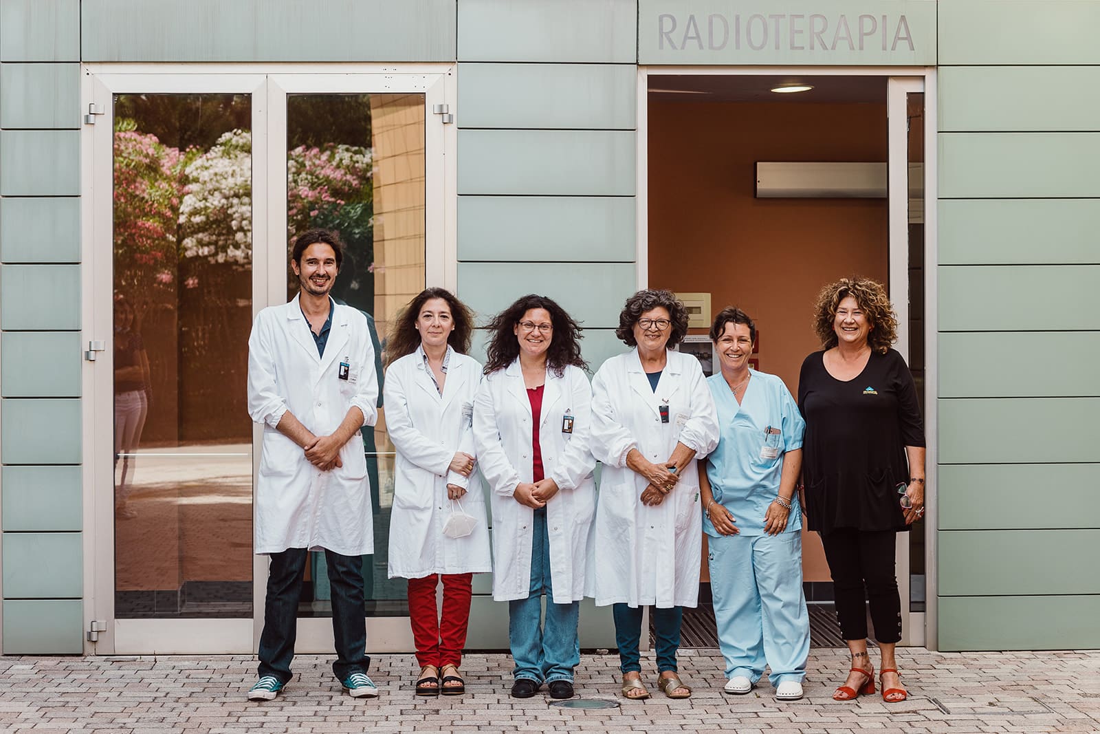
Radiotherapy at the Casa di Cura San Rossore has:
Computed tomography (Siemens Open Sensation CT), 64 multislice, entirely dedicated to radiation therapy
TrueBeam STx linear accelerator (see photo gallery above) from Varian Medical System, equipped with an integrated “cone beam CT” device that allows to perform, prior to treatment, a radiological evaluation thanks to which it is possible to monitor the real position of the tumor target and critical organs, correct any positioning errors, and evaluate any displacements even millimeters of the tumor target due to the often non-eliminable movement of internal organs ( organ motion ) and thus irradiate with greater precision.
The TrueBeam STx also has a micro-multilamellar system that allows for high-precision cranial and extracranial stereotactic treatments and, thanks to the machine’s ability to deliver X-radiation even in the FFF (Flattening Filter Free) mode, to decrease the time of dose delivery and thus of the treatment itself
Treatment planning systems (TPS) used in close collaboration with medical physicists
Image fusion systems using MiM system for CT, MRI and PET
Physical-dosimetric instrumentation used for periodic inspection of equipment
Immobilizers customized according to body district.
Casa di Cura San Rossore also has the following imaging equipment/technology for use in the execution of the treatment plan:
Internal: Magnetic Resonance Imaging 1.5 Tesla (from San Rossore Nursing Home Radiology)
External: PET and PET/CT in collaboration with the CNR Nuclear Medicine of Pisa and University Nuclear Medicine of the Azienda Univertsitaria-Ospedaliera S.Chiara of Pisa
This technological equipment then allows us to plan sophisticated radiotherapy treatments such as:
Volumetric Arc Radiotherapy ( VMAT)
Intensity Modulated Radiotherapy (IMRT )
Image-guided radiation therapy (IGRT)
3D-CRTconformal Radiotherapy
Intracranial and extracranial stereotactic radiotherapy (SRS, SBRT)
Adaptive Radiotherapy (ART)
Radiotherapy treatments with RESPIRATORY GATING widely used for breast neoplasms, lung etc.
RESPIRATORY GATING technique consists of a treatment in which irradiation is synchronized with the patient’s breathing with the intention of avoiding irradiation of healthy organs close to the tumor.
There are two ways to perform respiratory gating:
Free Breathing: the patient breathes freely and regularly, and beam delivery is performed in the respiratory phase where the tumor has reduced mobility. The progress of the respiratory cycle is continuously monitored during the treatment so as to make sure that dispensing is done at the correct phase of the cycle.
Deep inspiration breath hold (DIBH): in which the dispensing takes place during the patient’s apnea; for correct apnea amplitude and reproducibility, the patient has both during centering and treatment, an LED display that provides visual feedback of thoracic expansion correlated with respiratory movement, so as to help the patient immediately find the correct position (limiting the margins of error due to respiratory movements as much as possible
RADICAL-CURATIVE:
As an exclusive therapy:
– treatments with conventional fractionations
– hypofractionated treatments i.e., the ability to deliver high doses with the same or higher therapeutic efficacy thus reducing the overall duration of treatments compared to more traditional schemes
As combination radiotherapy, integrated in various modalities with surgery (preoperative, intraoperative, postoperative) or in combination with chemotherapy (neoadjuvant, concomitant, sequential), or in combination with hormone therapy or immunotherapy
PALLIATIVE-SYMPTOMATIC:To decrease and/or eliminate symptoms due to neoplastic disease such as pain , compressive syndromes (e.g. mediastinal syndrome, spinal cord compression, etc..) or rarely otherwise uncontrollable bleeding, and/or combined with other therapeutic weapons.
RADIOTHERAPY OF BENEFICIAL LESIONS:For example, pituitary adenomas, meningiomas, arteriovenous malformations (AVMs), Basedow’s disease, keloids, aneurysmal cysts, cranial nerve pathologies (e.g. trigeminal), vascular pathologies.
Clinical evaluation Oncology:
The first contact between the patient and the Center occurs with the first visit to the Radiotherapy Department (“Radiotherapy Oncology Consultation”). During the consultation, the radiation oncology oncologist collects all information, through medical records, about the patient’s current and remote health status to determine the nature and extent of the disease thus proceeding to multidisciplinary consultations.
Once the clinical issue is identified, the radiation oncologist analyzes the possibility of radiation treatment, choosing the most suitable delivery modality for the individual patient, in consultation with colleagues in Medical Physics.
Centering TC:
Upon accepting the patient for radiation therapy, the Radiation Oncologist and the Medical Radiology Health Technician (TSRM) perform a centering CT scan by defining the patient’s position on the couch and determining the type of immobilization system best suited for the type of therapy to be delivered.
Contornation:
Next, the radiation oncologist identifies the sensitive structures to be contoured on the centering CT scan. To do this it also uses information from other diagnostic investigations such as MRI and PET. Thanks to modern software in the Center, the radiation oncologist has the ability to “fuse” all the patient’s diagnostic images, managing to pinpoint with millimeter precision the region to be irradiated.
Development of the treatment plan:
The Medical Physicist prepares the Treatment Plan by optimizing the dose in the target volume while trying to spare the healthy organs surrounding the disease as much as possible. Each patient will therefore have a Care Plan that is strictly individualized, and suitable for achieving the most accurate dose distribution; consequently it will be different from that of any other patient. After the planning is finished, the Medical Physicist presents the plan to the Radiation Oncologist, which will be analyzed, discussed, possibly modified, and accepted.
Before being delivered to the patient, the Plan is verified on the ArcCheck dummy (for verification of rotational treatments) by evaluating its correct dose delivery (step in the overall Quality Control pathway)
Verification of positioning and treatment:
To carry out the treatment session, the patient is introduced inside the room (Bunker) where the linear accelerator is housed and is made to lie on the treatment couch. A CT scan is taken of the region to be irradiated before radiation is delivered and compared with the centering CT scan. With this procedure, it is possible to position the patient with pinpoint accuracy.The patient inside the therapy room is constantly monitored by the TSRM via a closed-circuit video system. In addition, an intercom system via intercom allows the patient to communicate with the technician. Throughout the radiotherapy treatment, the Patient will undergo follow-up visits during which any supportive therapy will be prescribed. Periodic follow-up visits are carried out after the conclusion of treatment.
Radiation treatment is performed either on an outpatient basis or on an inpatient or day-hospital basis for special clinical cases or for ‘performing combination therapies ( e.g., chemotherapy, nutrition, supportive medical therapy). Admission to the ward or dedicated housing is also possible for special logistical situations (Click here to see our solutions for your stay)
Radiation Therapy collaborates both within the Casa di Cura within dedicated Multidisciplinary Groups and with the various specialists at other National Centers for the discussion of the most complex clinical cases to ensure that each patient receives the proper integration and personalization of planned treatments based on International protocols and internal guidelines adapted to the clinical situation of each individual patient.
The Physician verifies and evaluates all parameters of your treatment plan, verifies every radiological image taken before and during treatment including by Cone Beam verifications at the time of treatment for correctness of therapy; performs intercura
The medical physicist:
It ensures through periodic checks that the characteristics of the equipment are within the tolerance levels chosen to ensure the optimal performance of treatments. It ensures the treatment pathway: from the preparation of the treatment plan, to its approval following discussion with the physician, from the transfer of the plan to the treatment machine through the Record and Verify (ARIA) system that allows the identification of the patient, the correct association of his or her treatment plan and the recording of the dose delivered to each fraction, to the verification before the start of the treatment of the correct delivery of the plan by the equipment and also supervises with other working figures (physicians and technicians) during treatments with respiratory gating.
The TSRM health technician:
Works according to the instructions of the radiation oncologist at the various stages of the radiation therapy procedure: performs patient set up, if necessary using positioning and immobilization systems; during the simulation phase of the treatment, acquires the images necessary for the production of the treatment plan and prepares the accessories necessary for its implementation (shielding, etc.); performs the radiation treatment according to the indications of the plan prepared and approved by the physicist and physician and is responsible for the correct application of the plan itself; acquires the images produced with IGRT technology and assists the radiation oncologist in the “on line” control for the correction of the set up. Prior to the start of treatment carry out the check together with the physicist of the correct delivery of the plan by the machine.
Dr. Maria Grazia Fabrini – Head of the Radiotherapy Operating Unit
Dr. Francesco Cartei – Radiotherapy
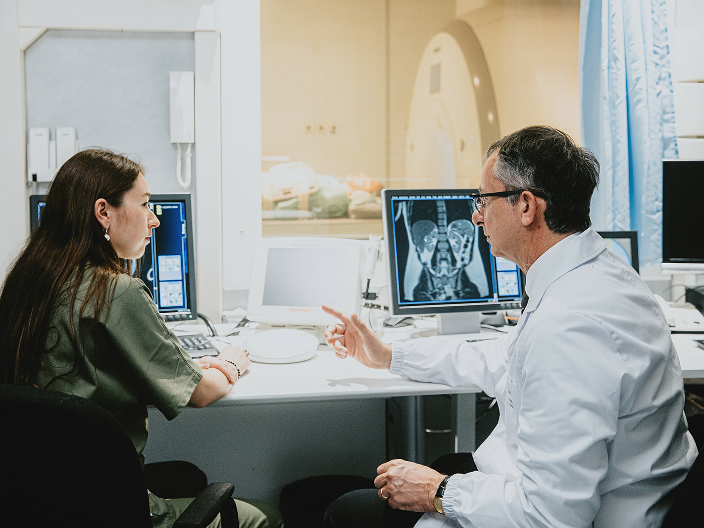
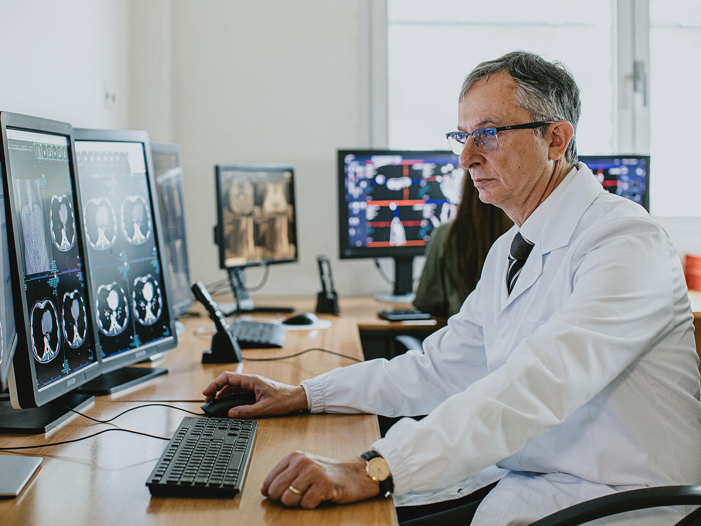
Radiodiagnostics
Radiodiagnostics
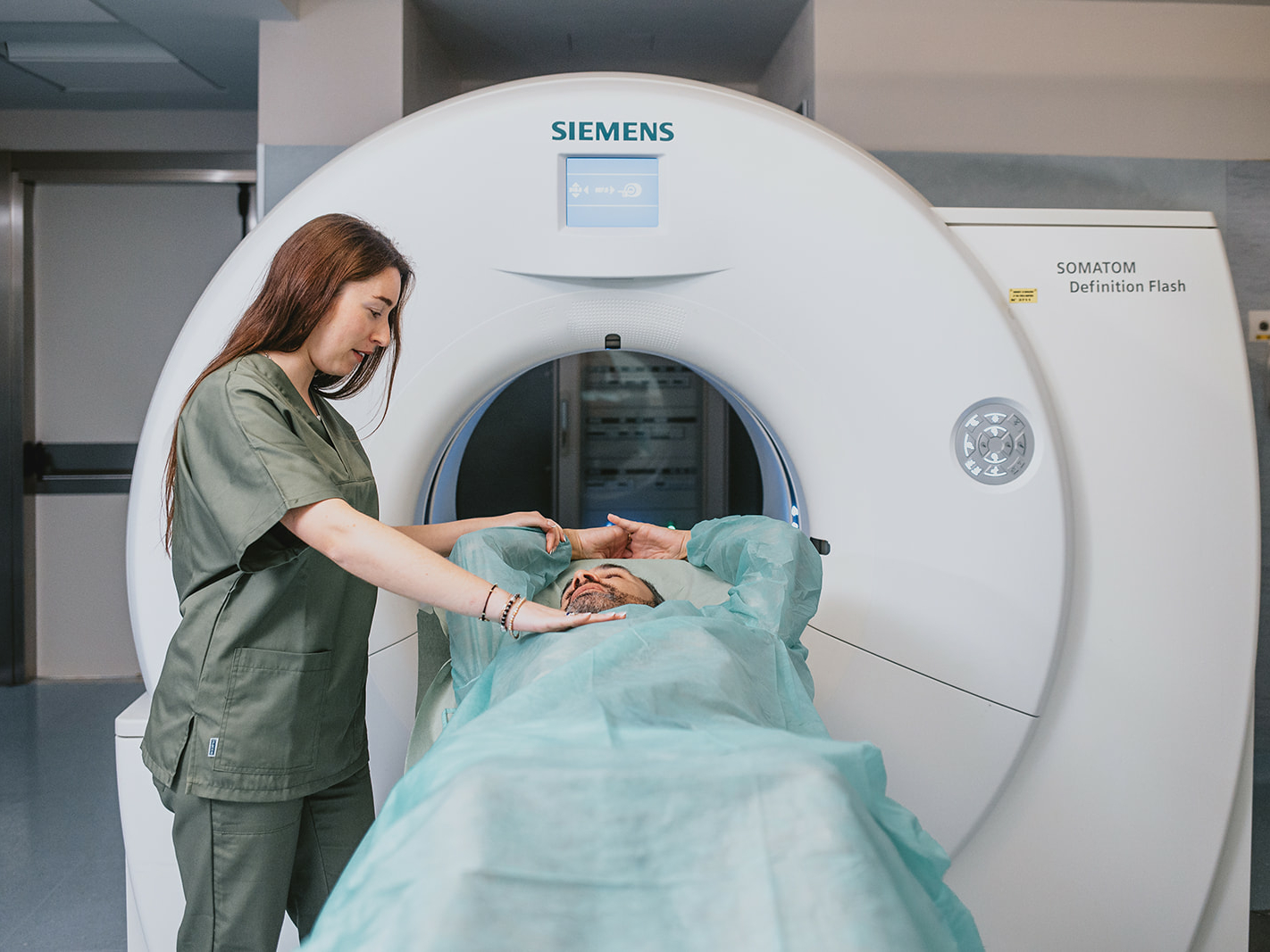
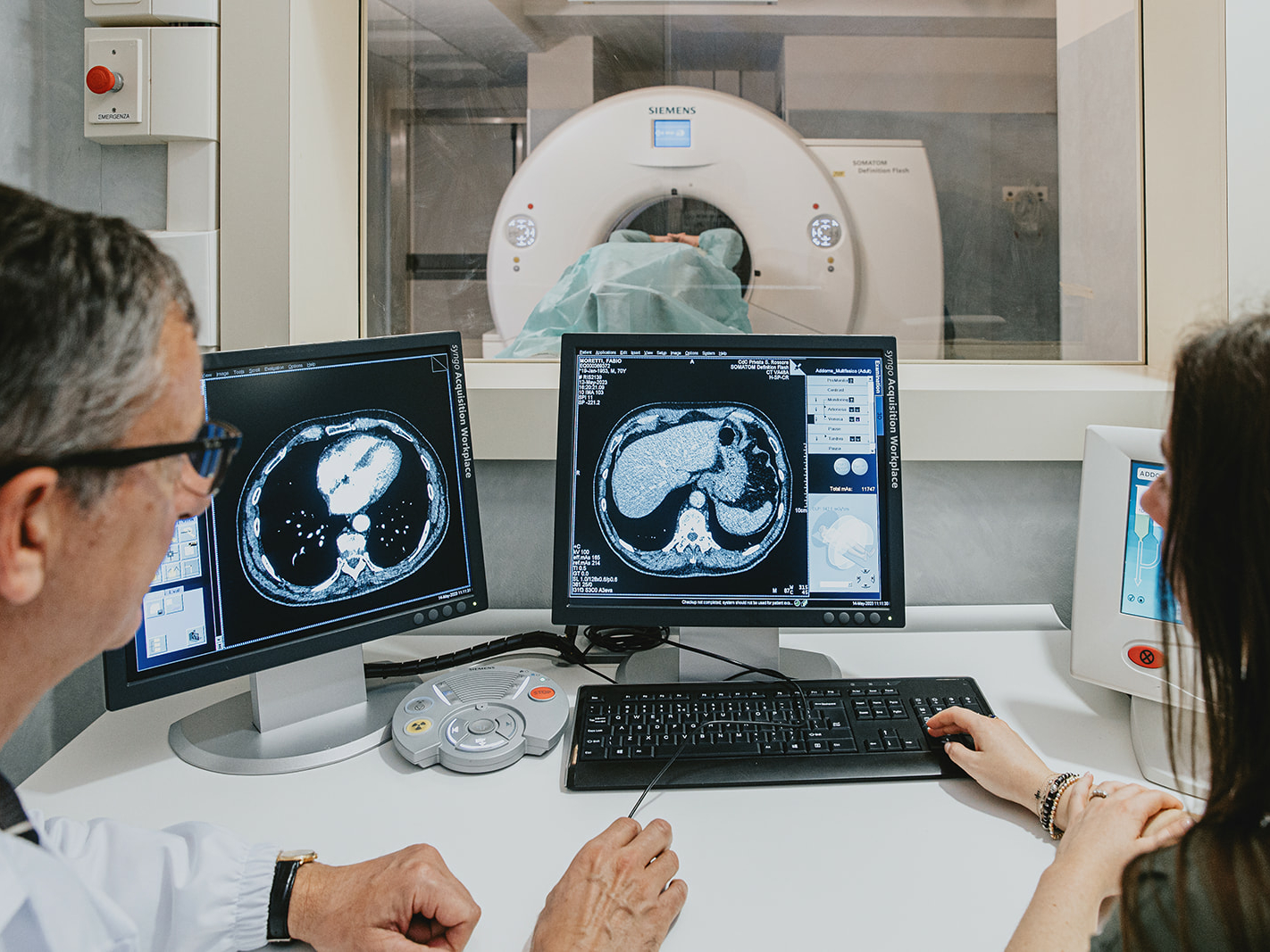
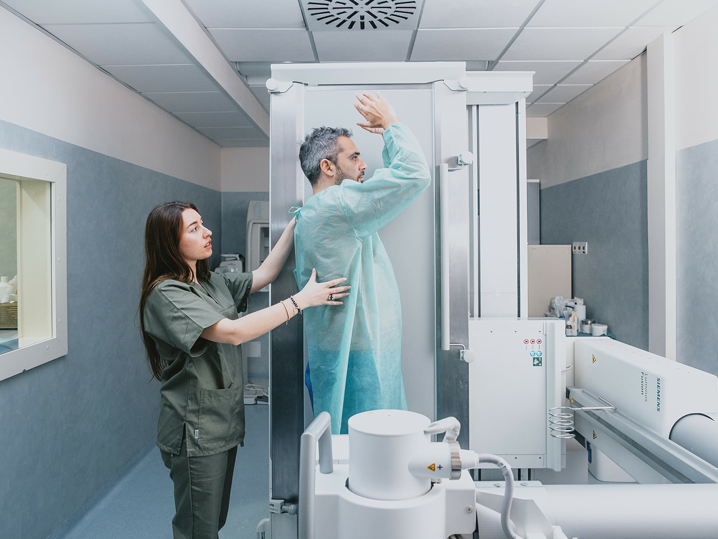
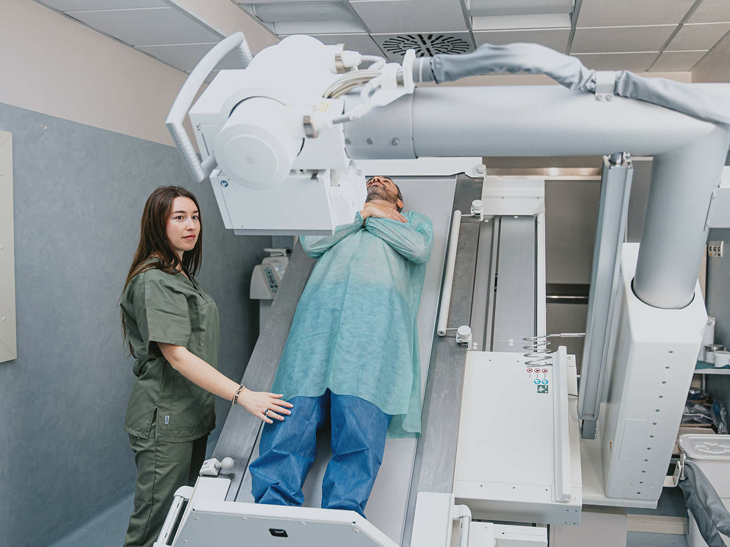
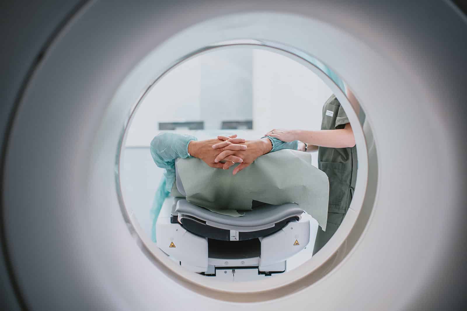
At the Casa di Cura San Rossore it is possible to perform diagnostic tests and therapeutic treatments, even without the need for hospitalization.
Our center employs qualified doctors and state-of-the-art machinery: medical personnel are selected based on parameters such as skills, qualifications and experience in the field.
The center’s wide opening hours meet the needs of all patients; in addition, thanks to the special platform, reports can be viewed quickly and easily at any time.
Rx joint districts
Rx esophagus-stomach-duodenum with MDC
Rx clisma opaque with MDC
Rx defecography with MDC
Rx urethrocystography/ cystoscopy with MDC
Rx abdomen
Chest Rx
Rm peripheral joint districts with and without MDC
Skull Rm with and without MDC
Rm neck with and without MDC
Rm abdomen with and without MDC
Cardiac MRI with and without MDC
Rm spine
Multiparametric prostatic Rm
Massive facial MRI with and without contrast medium
Rm colangium
Tc peripheral joint districts
Tc spine
Tc abdomen with and without MDC
Chest CT with and without MDC
Skull Tc with and without MDC
Total body Tc with and without MDC
Coronary Tc with and without MDC
Tc dentalscan dental arches
Virtual colonoscopy tc
Dr. Alessandro Paolicchi – Radiology service manager, specialist in diagnostic imaging
Dr. Alessandro Lunardi – Interventional radiology
Dr. Mario Sabato – Radiodiagnostics
Dr. Anna Cilotti – Breast Radiodiagnostics
Dr. Carolina Marini – Breast Radiodiagnostics
Dr. Barbara Ginanni – Breast Radiodiagnostics
Dr. Massimo Lombardi – Radiologist Cardiologist
Dr. Salvatore Mazzeo – Radiologist Endocrinologist
Dr. Cristina Bisogni – Radiodiagnostics
INFO POINT
REQUEST INFORMATION
For questions, clarifications or other Info please fill out the form below, you will be contacted by our dedicated staff as soon as possible.
Pathological anatomy
Pathological anatomy
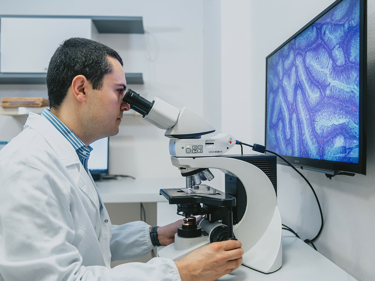
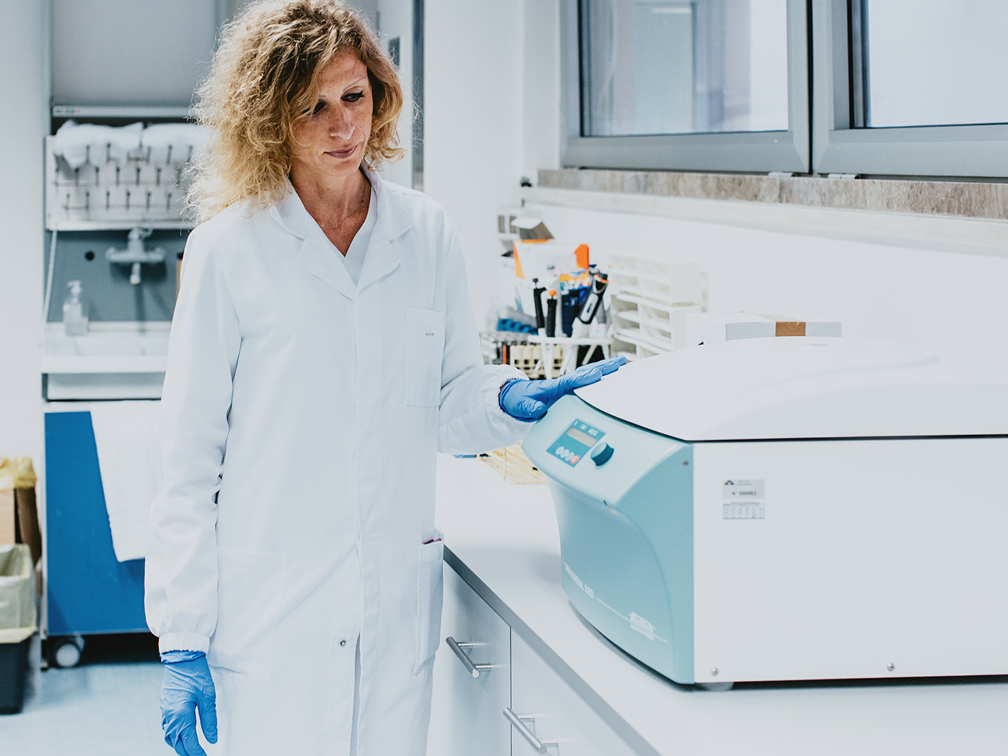
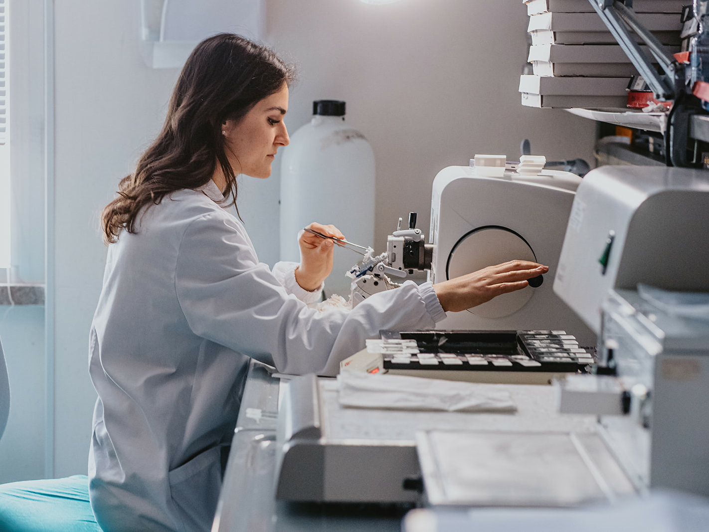
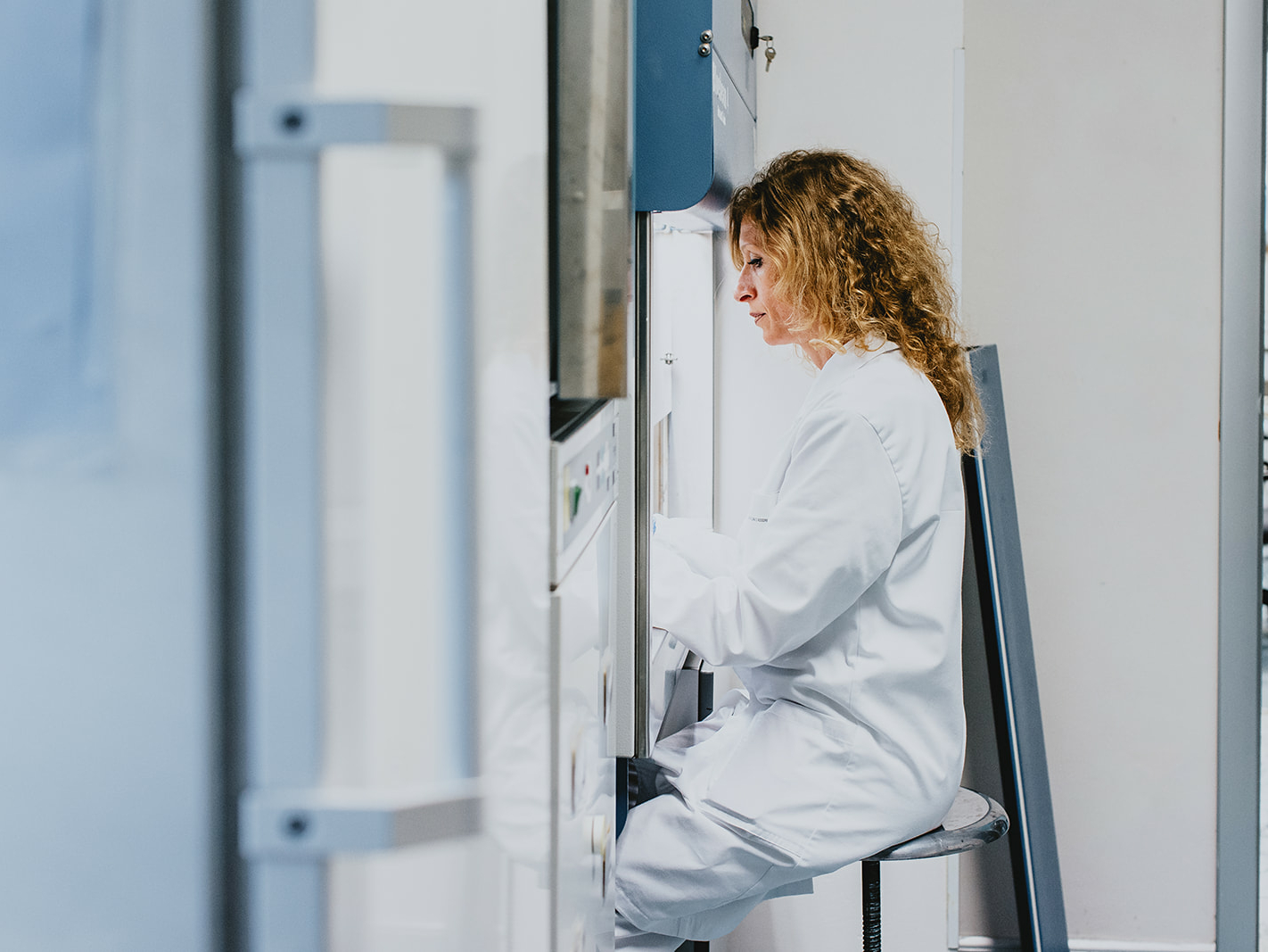
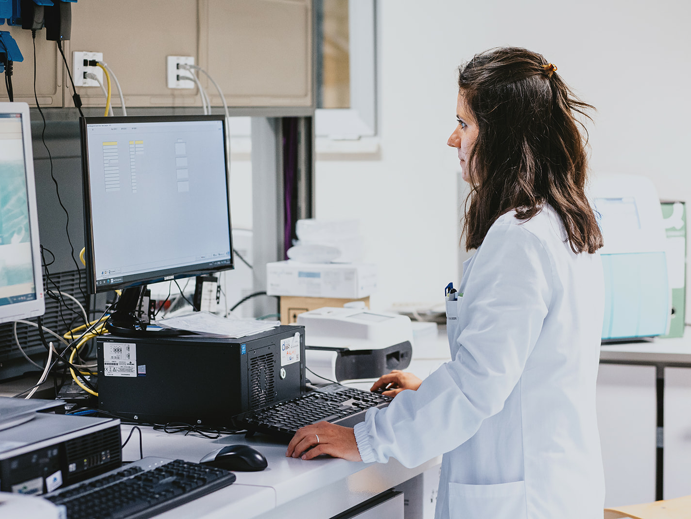
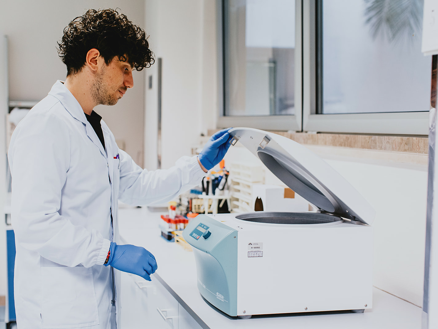
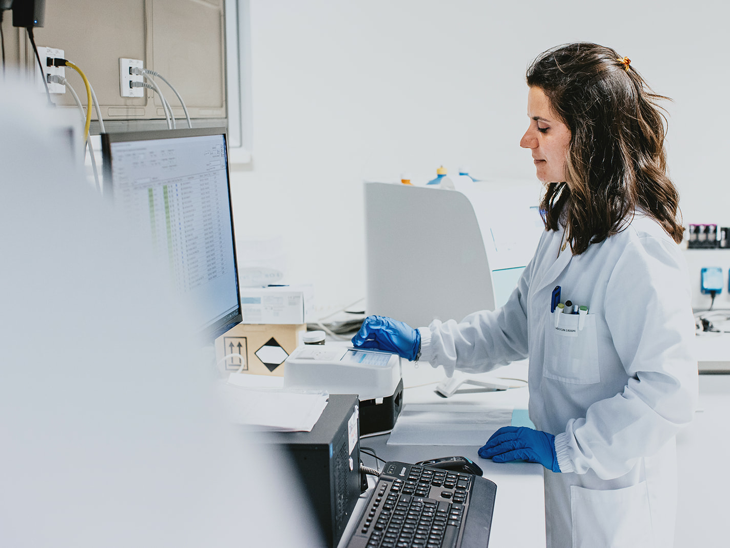
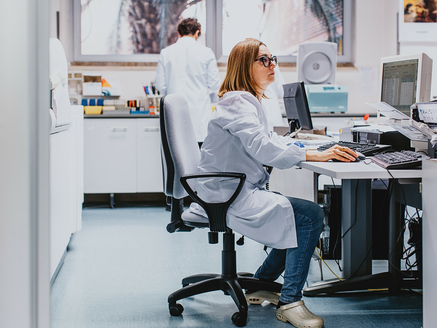
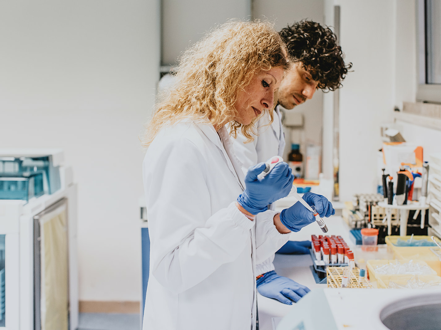
The Laboratory Medicine Area of the Casa di Cura San Rossore is divided into two Sections:
–Chemical-Clinical Analysis and Microbiology
– Pathological Anatomy: Histology, Clinical Cytology, Cervicovaginal Cytology accompanied by HPV molecular research and characterization, Molecular Diagnostics of Neoplasms
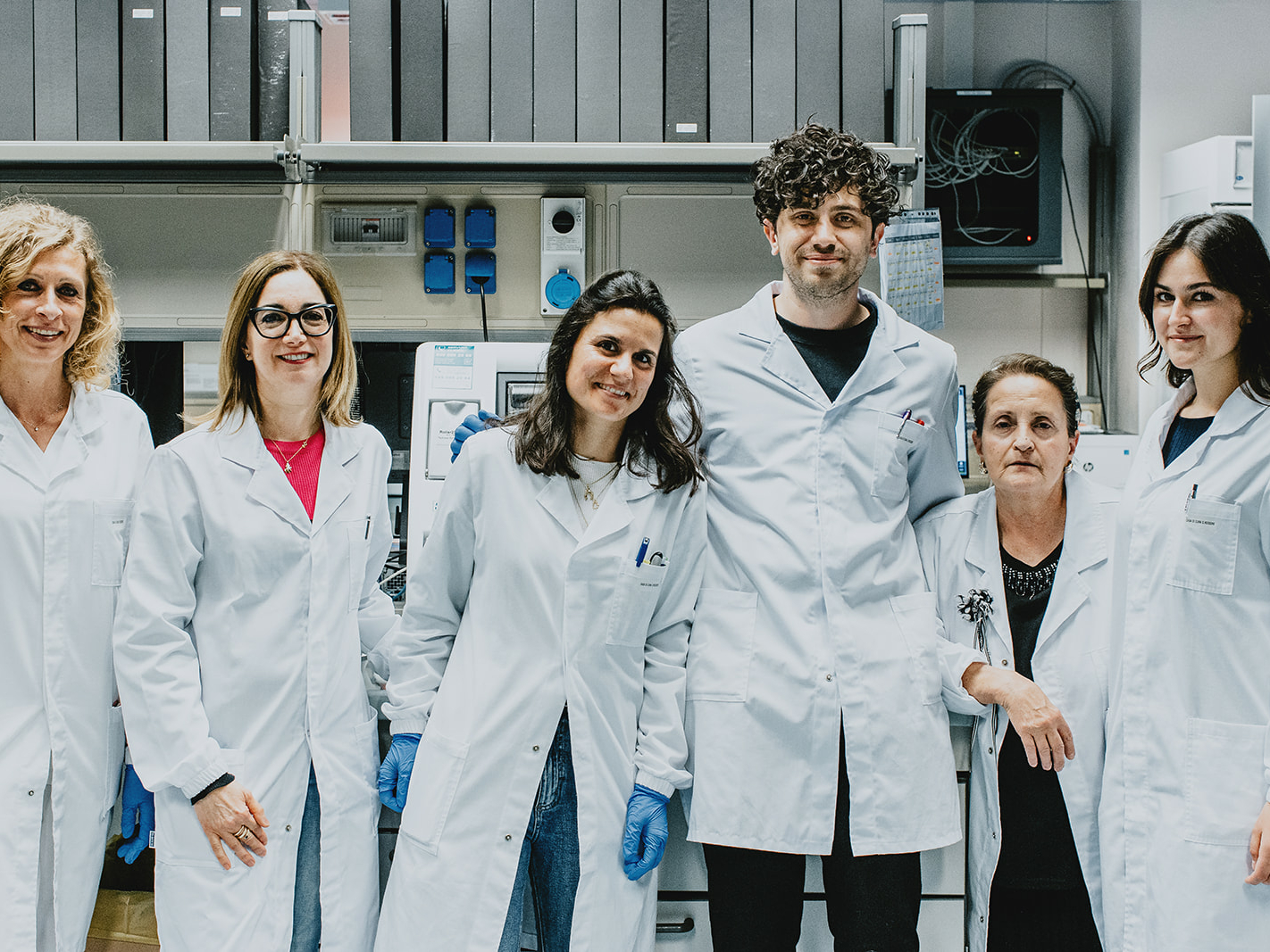
The Laboratory supports the activities carried out within the Casa di Cura San Rossore for both outpatients and inpatients. In addition, the Laboratory is also available for examination requests from outside.
The Laboratory is open 24 hours a day for inpatient activity needs and provides the histo-cytopathological diagnosis in a very short time: same-day for clinical cytology, 48 hours for biopsies, 72 hours for surgical material.
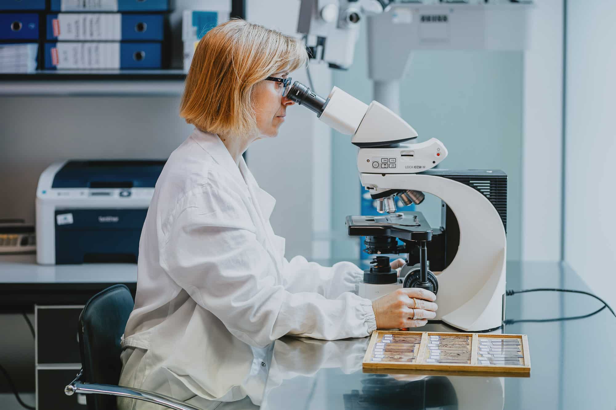
Dr. Valerio Ortenzi – Director of the Department of Pathological Anatomy
Dr. Francesca Garbini – Specialist in Pathologic Anatomy
Endoscopy
Endoscopy
Casa di Cura performs Endoscopy examinations for diagnostics, therapeutic treatment, and the performance of and possible support for surgical procedures.
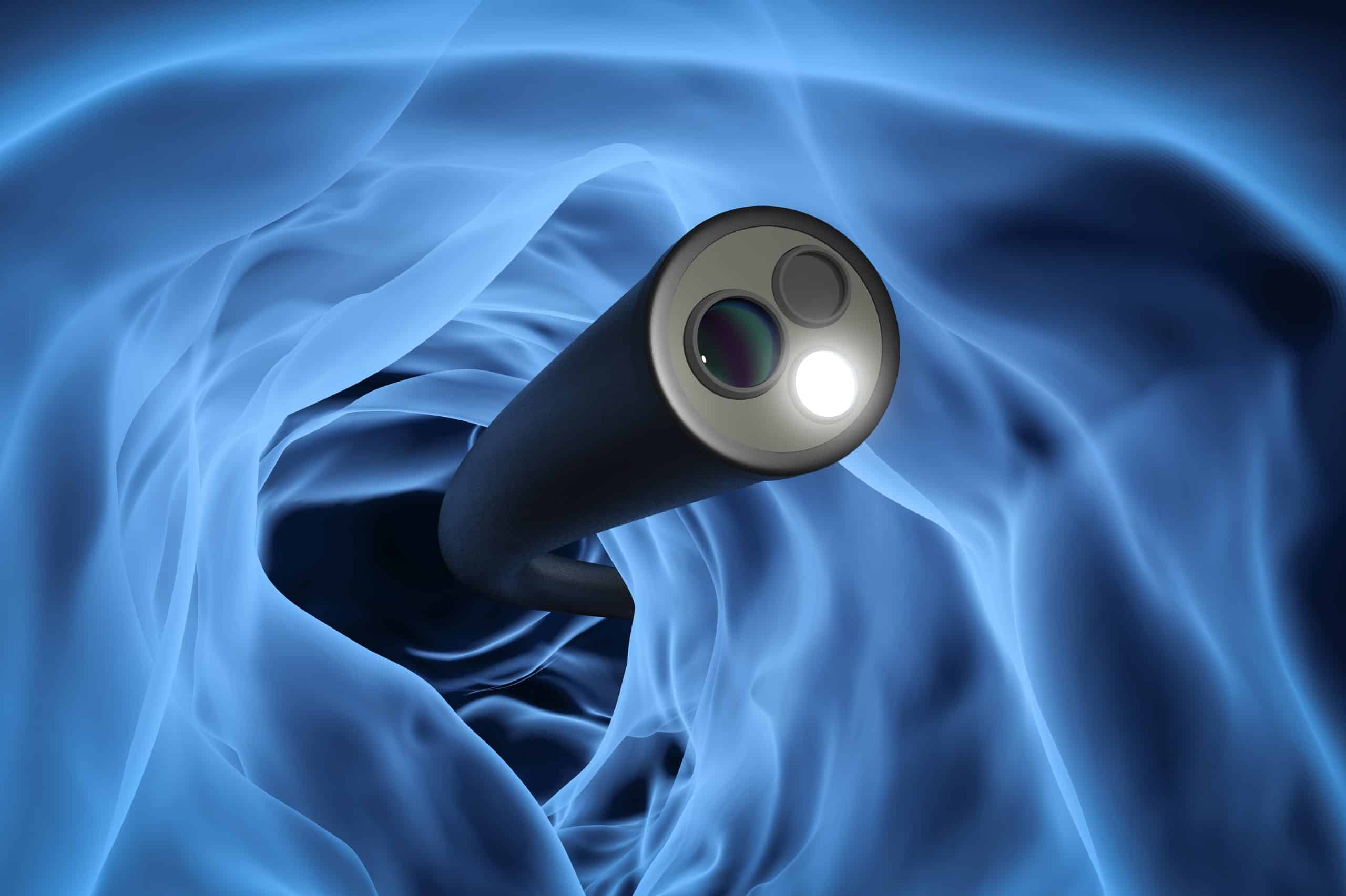
The examinations, performed if necessary with anesthesiological assistance, are:
Esophago-gastro-duodenoscopy (Gastroscopy)
Pancolonoscopy (Colonoscopy)
Vital coloring
Endoscopic magnification
Esophageal pH-metry (conventional or with wireless capsule)
High-resolution esophageal manometry
Rigid dilatation, hydraulic, pneumatic
Placement PEG (Percutaneous Endoscopic Gastrostomy)
Endoprosthesis placement
Mucosectomia (EMR, Endoscopic Mucosal Resection)
Endoscopic submucosal resection (ESD)
Radiofrequency Ablation (RFA)
Treatment of esophageal fistulas and perforations
Endoscopic myotomy for achalasia (P.O.E.M., PerOral Endoscopic Myotomy)
Video-capsule endoscopy allows visualization of the colorectal mucosa by ingesting a small capsule-shaped camera that, as it advances along the digestive tract, records images and sends them to a small external recorder strapped to a belt that the patient wears around the waist.Compared with traditional colonoscopy and virtual colonoscopy, video-capsule colonoscopy allows direct visualization of the colon mucosa (like the traditional exam), does not rely on radiation (like virtual colonoscopy), and most importantly is non-invasive (but not therapeutic).
Dr. Giovanni Pallabazzer – General Surgeon, Endoscopist, perfected in the diagnosis and treatment of diseases of the esophagus and stomach
Dr. Lorenzo Bertani – Gastroenterologist
Dr. Adolfo G. Puglisi – General surgeon, specializing in Endoscopy and Proctology
Dr. Biagio Solito – General Surgeon, specializing in Esophageal Surgery, Endoscopy and Proctology
Dr. Roberto Spinsi – General surgeon, specializing in endoscopy of the digestive tract
Dr. Alessandro Sturiale – Colo-proctology surgeon, specializing in pelvic floor surgery
Dr. Gianluca Toniolo – Specialist in Surgery, Endoscopy and Anesthesiology
Laboratory Examinations
Laboratory Examinations
The Laboratory Medicine Area of Casa di Cura San Rossore is divided into two sections:
–Chemical-Clinical Analysis and Microbiology
– Pathological Anatomy: Histology, Clinical Cytology, Cervicovaginal Cytology accompanied by HPV molecular research and characterization, Molecular Diagnostics of Neoplasms
The Laboratory supports the activities carried out within the Casa di Cura San Rossore for both outpatients and inpatients. In addition, the Laboratory is also available for examination requests from outside.
The Laboratory is open 24 hours a day for inpatient activity needs and provides the histo-cytopathological diagnosis in a very short time: same-day for clinical cytology, 48 hours for biopsies, 72 hours for surgical material.
Responsible
Dr. Valerio Ortenzi
Pathological Anatomy
Dr. Francesca Garbini
